Project Description
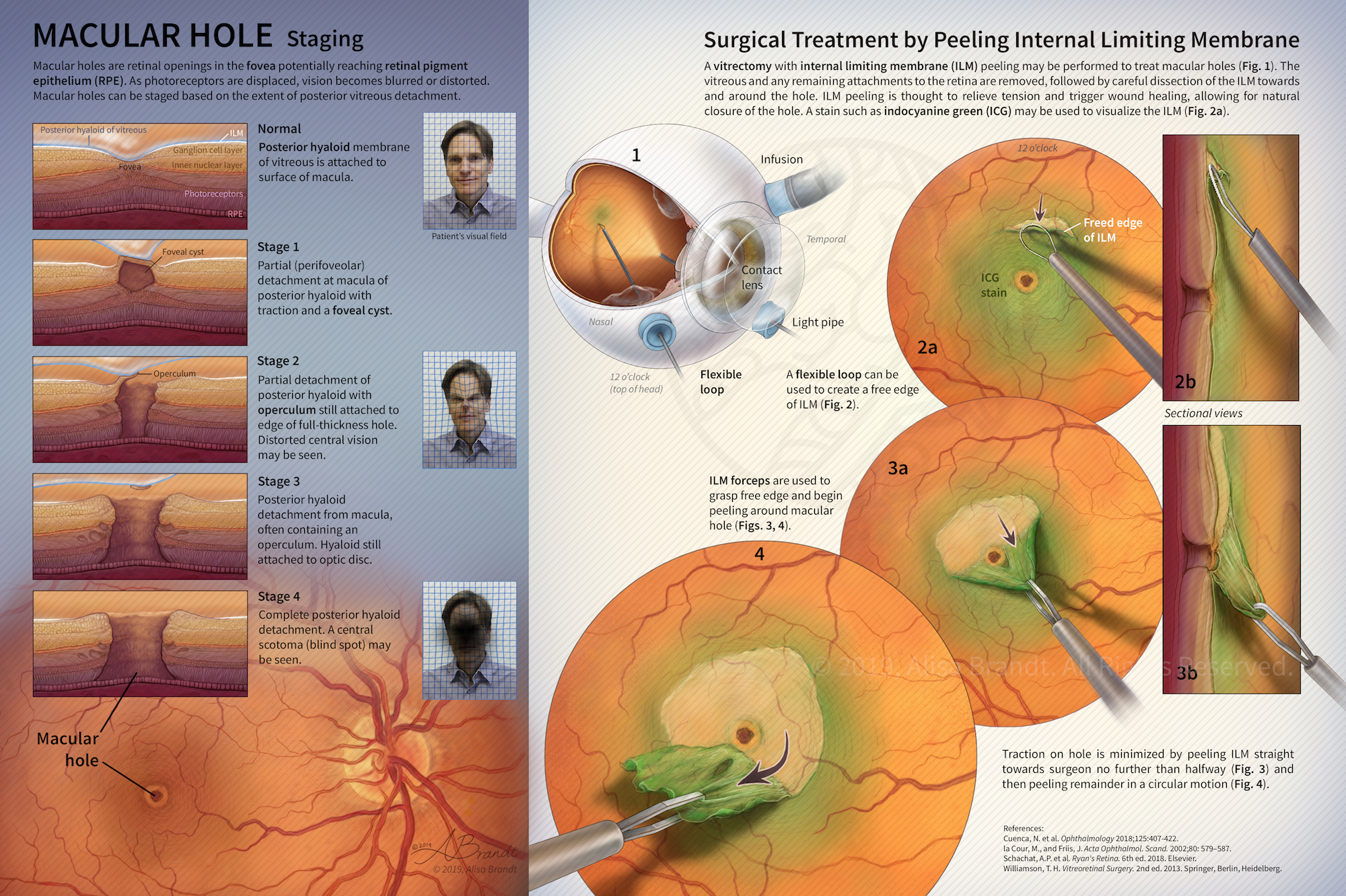
Illustration explaining stages in macular hole formation in the fovea and surgical treatment by peeling the internal limiting membrane during a vitrectomy procedure. Figure 1 and surgical instruments were first created in ZBrush or Cinema 4D, followed by painting in Photoshop. Designed to be a fold-out page in a scientific or medical journal. Created during the Ophthalmological Illustration course in the Medical and Biological Illustration graduate program at Johns Hopkins University School of Medicine.