
Hipbone
Hipbone in carbon dust from direct observation, created during the Medical and Biological Illustration graduate program at Johns Hopkins University School of Medicine.
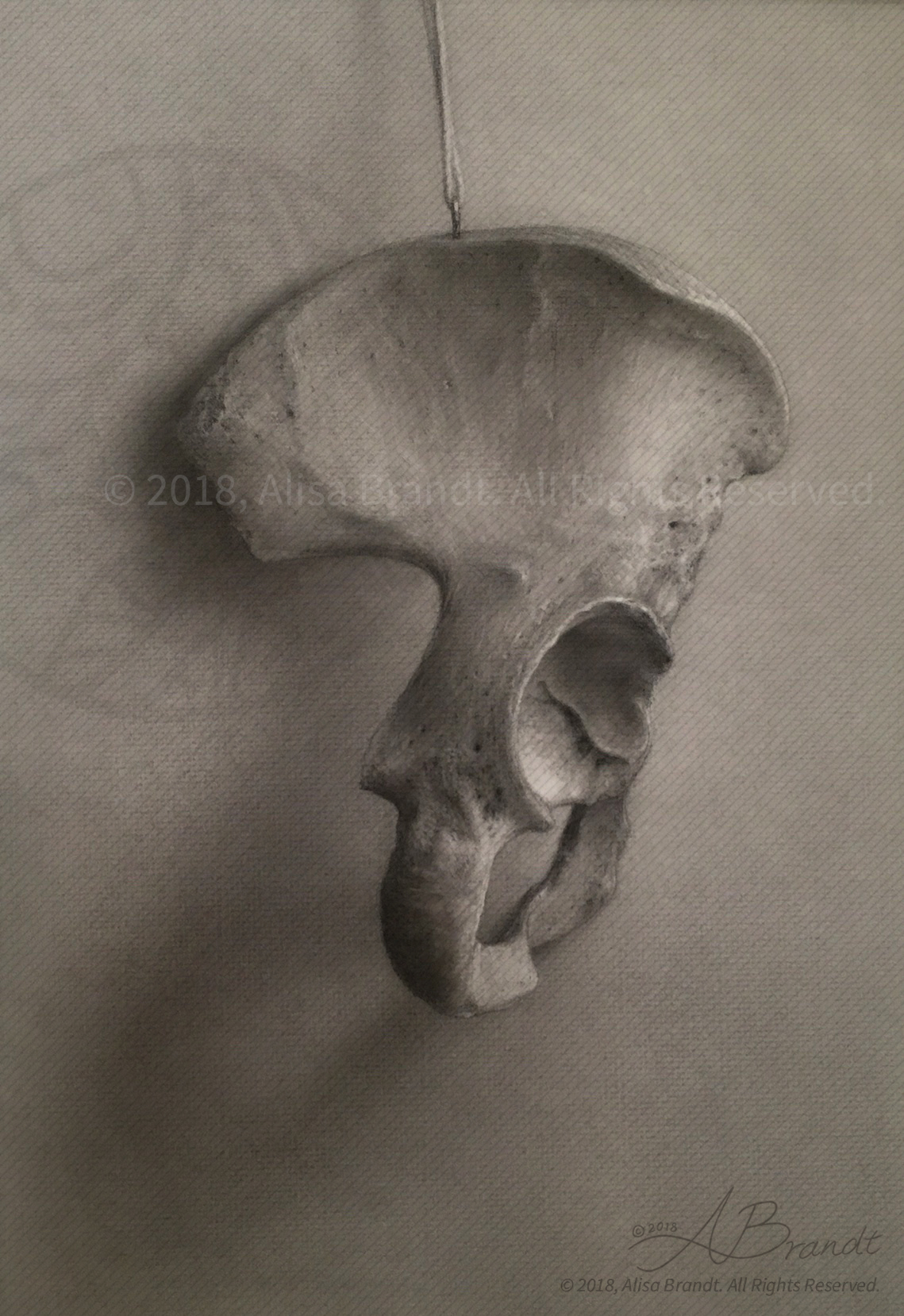
Hipbone in carbon dust from direct observation, created during the Medical and Biological Illustration graduate program at Johns Hopkins University School of Medicine.

Illustration created during the Neuroanatomy course in the Medical and Biological Illustration graduate program at the Department of Art as Applied to Medicine, Johns Hopkins University School of Medicine. This illustration teaches the anatomy of the human brain around the lateral ventricle, a fluid-filled space that extends into the temporal lobe. The brain has been cut in several planes to expose the floor of the left lateral ventricle and the