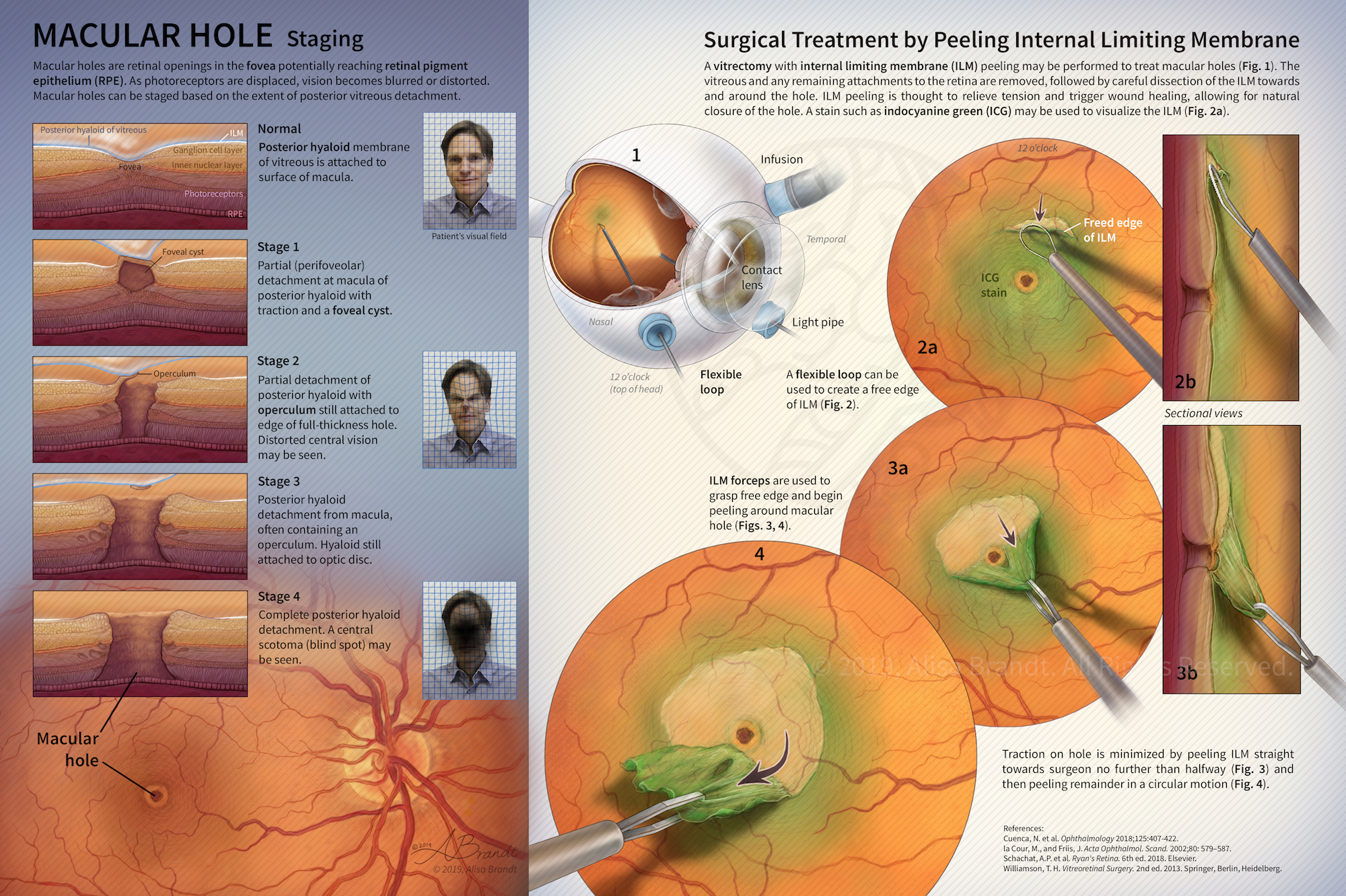
Macular hole staging and surgical treatment illustration © 2019 Alisa Brandt

Macular Hole: Staging and Surgical Treatment

Illustration explaining stages in macular hole formation in the fovea and surgical treatment by peeling the internal limiting membrane during a vitrectomy procedure. Figure 1 and surgical instruments were first created in ZBrush or Cinema 4D, followed by painting in Photoshop. Designed to be a fold-out page in a scientific or medical journal. Created during the Ophthalmological Illustration course in the Medical and Biological Illustration graduate program at Johns Hopkins University

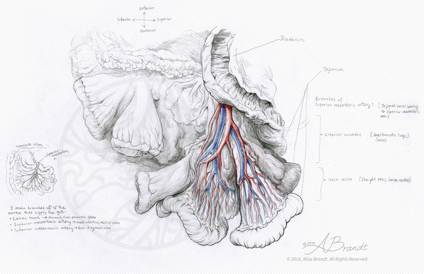
Anatomical sketch of mesentery

Small Intestine Mesentery (sketch)

Anatomical sketch from direct observation of the mesentery and mesenteric vessels of the small intestine. Created during the Medical and Biological Illustration graduate program at Johns Hopkins University School of Medicine.
Bumblebees poster

Nectar and Pollen Gathering of Bumblebees

Educational poster about bumblebees and their adaptations for nectar and pollen gathering, combining graphite and Photoshop. Created during the Medical and Biological Illustration graduate program at Johns Hopkins University School of Medicine.

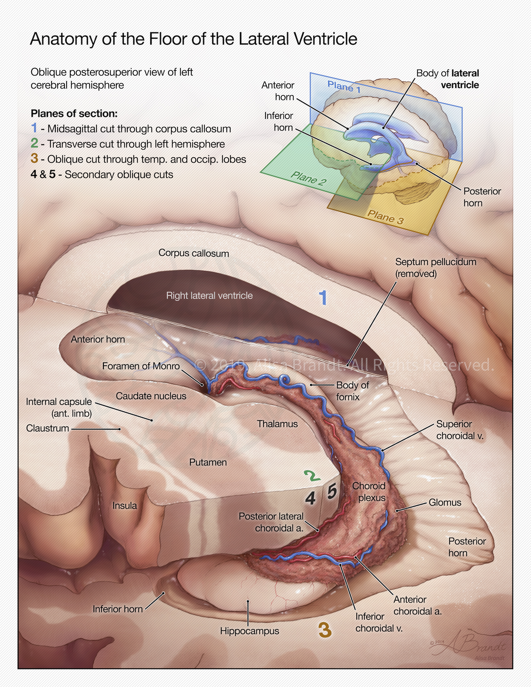
Neuroanatomical illustration

Anatomy of the Floor of the Lateral Ventricle

Illustration created during the Neuroanatomy course in the Medical and Biological Illustration graduate program at the Department of Art as Applied to Medicine, Johns Hopkins University School of Medicine. This illustration teaches the anatomy of the human brain around the lateral ventricle, a fluid-filled space that extends into the temporal lobe. The brain has been cut in several planes to expose the floor of the left lateral ventricle and the

Scroll UpScroll Up
error: Content is protected !!