Macular hole staging and surgical treatment illustration © 2019 Alisa Brandt

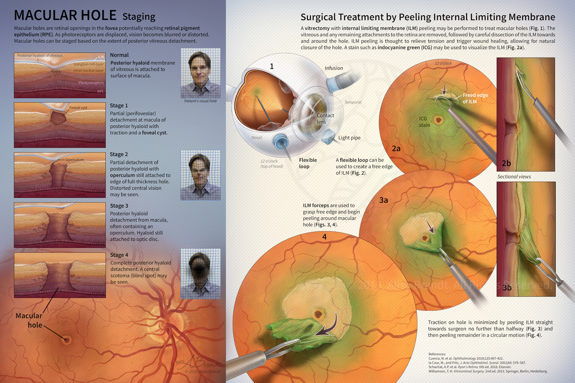
Macular Hole: Staging and Surgical Treatment

Illustration explaining stages in macular hole formation in the fovea and surgical treatment by peeling the internal limiting membrane during a vitrectomy procedure. Figure 1 and surgical instruments were first created in ZBrush or Cinema 4D, followed by painting in Photoshop. Designed to be a fold-out page in a scientific or medical journal. Created during the Ophthalmological Illustration course in the Medical and Biological Illustration graduate program at Johns Hopkins University

Still images from inner and middle ear turntable movie

The Inner and Middle Ear (stills)

Still images from a turntable movie (click here) of the inner and middle ear sculpted using ZBrush, based on MRM (magnetic resonance microscopy) data. The anatomy of the inner and middle ear is presented with a particular focus on the nerves of the area. The MRM data (a series of TIFF images) was prepared in OsiriX and a 3D surface was exported to OBJ format for sculpting in ZBrush. Data

Scroll UpScroll Up
error: Content is protected !!