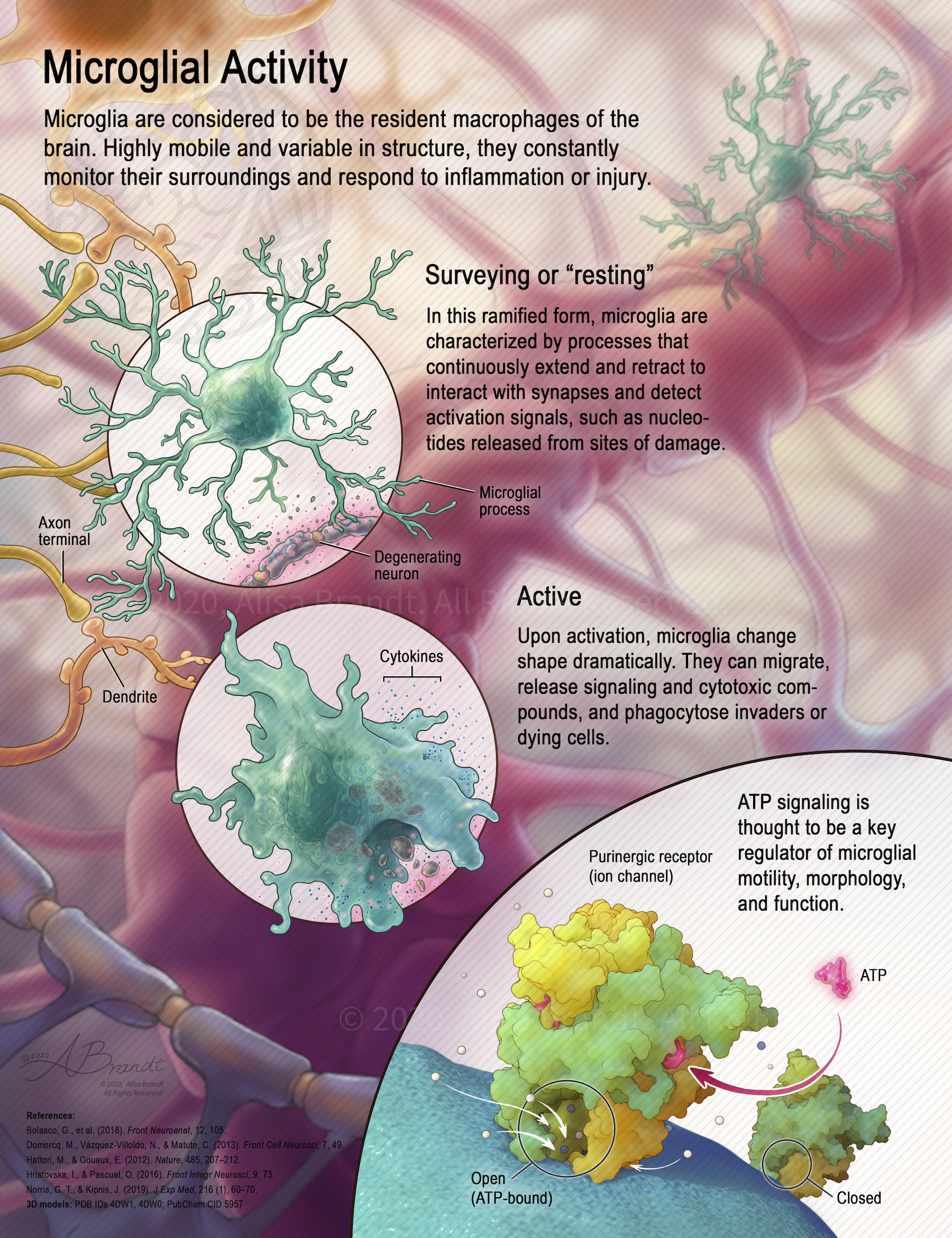
Neuroscience/cellular illustration about microglia © 2020 Alisa Brandt

Microglial Activity

This illustration is intended to provide neuroscience students an overview of specialized brain cells called microglia, by distilling complex, growing research into a concise and memorable poster. A defining characteristic of microglia is their ability to transform dramatically and take on different shapes and functions to monitor the neuronal environment and respond quickly to problems. Two main states are depicted in this illustration: a highly-branched “surveying” form, and an “active”

Macular hole staging and surgical treatment illustration © 2019 Alisa Brandt

Macular Hole: Staging and Surgical Treatment

Illustration explaining stages in macular hole formation in the fovea and surgical treatment by peeling the internal limiting membrane during a vitrectomy procedure. Figure 1 and surgical instruments were first created in ZBrush or Cinema 4D, followed by painting in Photoshop. Designed to be a fold-out page in a scientific or medical journal. Created during the Ophthalmological Illustration course in the Medical and Biological Illustration graduate program at Johns Hopkins University

Anatomical illustration of right eye cross-section © 2019 Alisa Brandt

Anatomy of the Human Eye

Anatomical illustration of a right human eye in horizontal cross-section. Measurements and OCT (optical coherence tomography) images were used as reference to create an accurate drawing. Created during the Ophthalmological Illustration course in the Medical and Biological Illustration graduate program at Johns Hopkins University School of Medicine.

Tissue painting using photo reference

Tissue Painting

Photoshop painting of various tissues, from photo reference. Created during the Medical and Biological Illustration graduate program at Johns Hopkins University School of Medicine.

Left frontal craniotomy surgical illustration

Left Frontal Craniotomy for Tumor Resection

Illustration of a left frontal craniotomy to remove a large brain tumor, designed for slide presentation. Created during the Surgical Illustration course in the Medical and Biological Illustration graduate program at Johns Hopkins University School of Medicine.

Superficial parotidectomy surgical illustration

Superficial Parotidectomy

Illustration of tumor removal from the parotid gland, designed for a textbook. Created during the Surgical Illustration course in the Medical and Biological Illustration graduate program at Johns Hopkins University School of Medicine.

Red-capped cardinals poster

Color Change in Maturing Red-Capped Cardinals

Illustration created for the National Aquarium in Baltimore, Maryland, during the Medical and Biological Illustration graduate program at Johns Hopkins University School of Medicine. This poster communicates the typical progression of color change in red-capped cardinals as they mature from fledgling to adult.

Bumblebees poster

Nectar and Pollen Gathering of Bumblebees

Educational poster about bumblebees and their adaptations for nectar and pollen gathering, combining graphite and Photoshop. Created during the Medical and Biological Illustration graduate program at Johns Hopkins University School of Medicine.

Laparoscopic sigmoid colectomy surgical illustration

Laparoscopic Sigmoid Colectomy

Laparoscopic sigmoid colectomy surgical illustration designed for a surgical atlas. Created during the Surgical Illustration course in the Medical and Biological Illustration graduate program at Johns Hopkins University School of Medicine.

Scroll UpScroll Up
error: Content is protected !!