
External Eye
The external eye in watercolor from direct observation in a mirror. Created during the Ophthalmological Illustration course in the Medical and Biological Illustration graduate program at Johns Hopkins University School of Medicine.
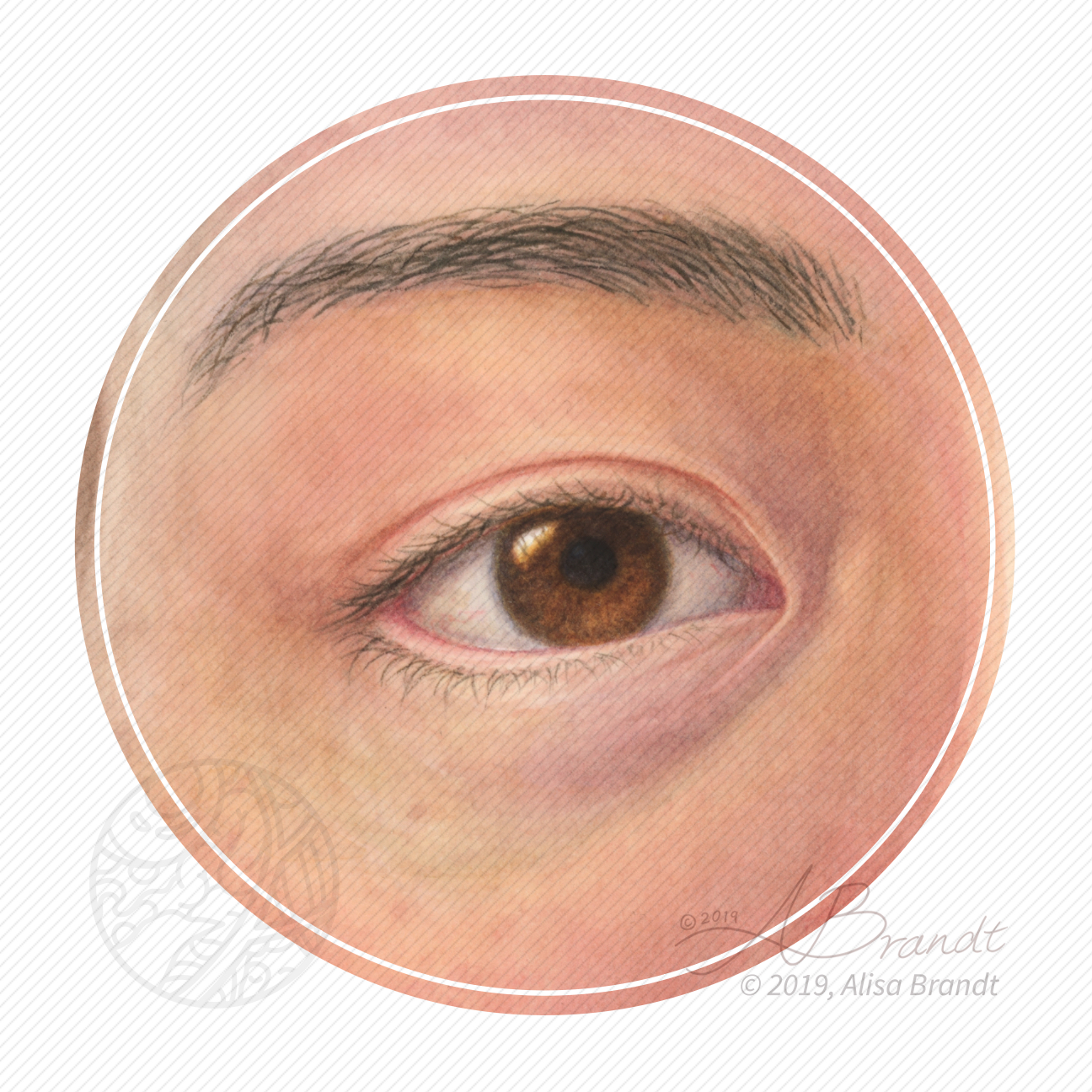
The external eye in watercolor from direct observation in a mirror. Created during the Ophthalmological Illustration course in the Medical and Biological Illustration graduate program at Johns Hopkins University School of Medicine.
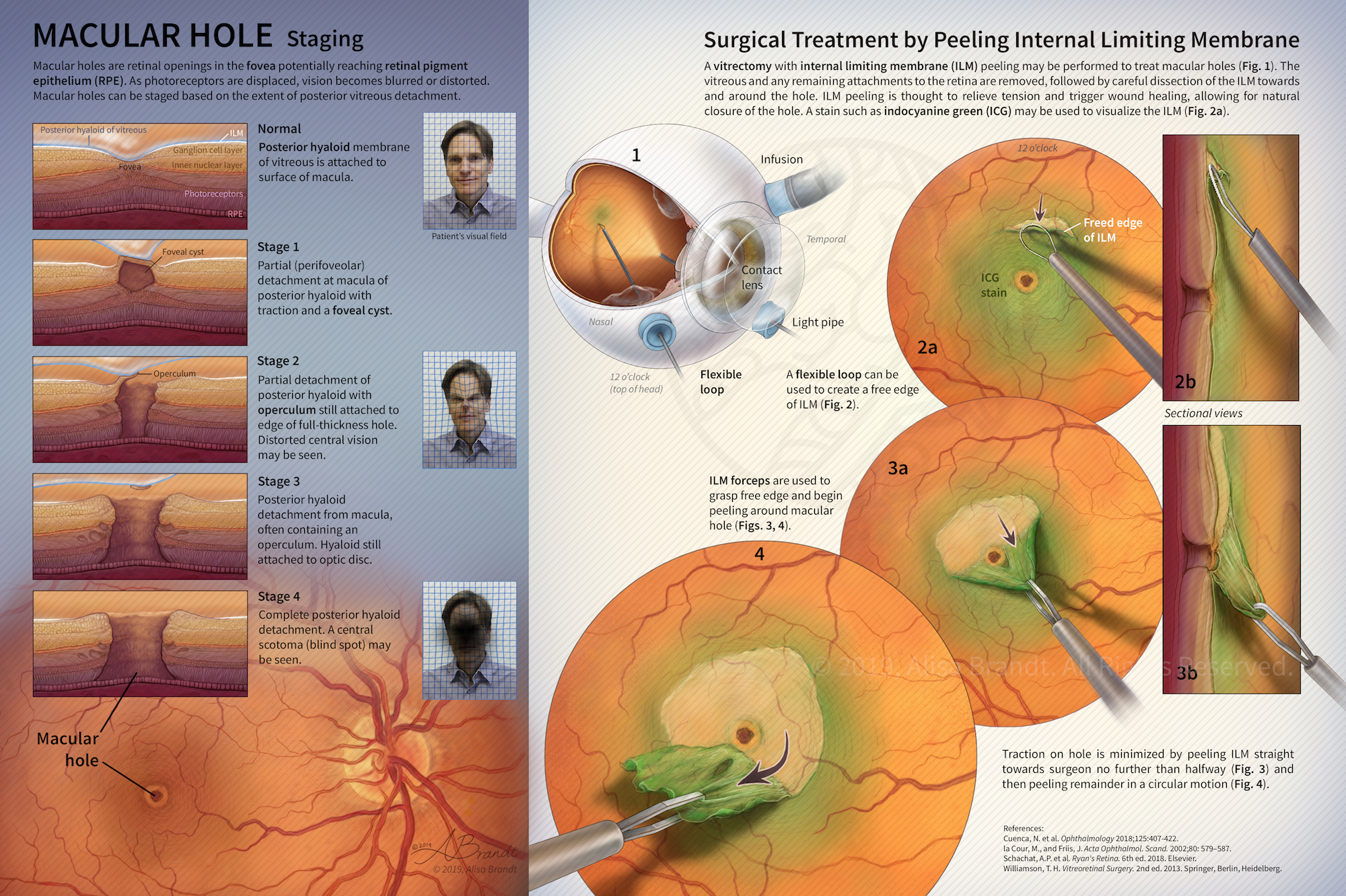
Illustration explaining stages in macular hole formation in the fovea and surgical treatment by peeling the internal limiting membrane during a vitrectomy procedure. Figure 1 and surgical instruments were first created in ZBrush or Cinema 4D, followed by painting in Photoshop. Designed to be a fold-out page in a scientific or medical journal. Created during the Ophthalmological Illustration course in the Medical and Biological Illustration graduate program at Johns Hopkins University
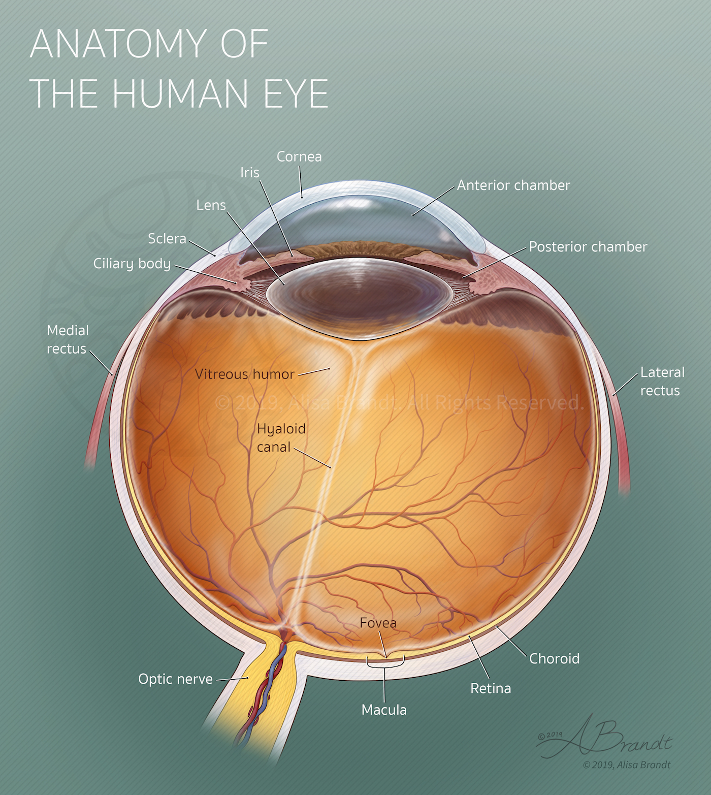
Anatomical illustration of a right human eye in horizontal cross-section. Measurements and OCT (optical coherence tomography) images were used as reference to create an accurate drawing. Created during the Ophthalmological Illustration course in the Medical and Biological Illustration graduate program at Johns Hopkins University School of Medicine.

Team project to design and propose a mock mobile application that functions as both an interactive sleep journal and a resource for learning about sleep and common sleep disorders. Target audience members were adults experiencing symptoms of sleep disorders who are interested in building a self-directed record of their sleep and learning about sleep health. A 50-page proposal booklet was created containing background research, a style sheet, flowcharts, storyboards, and
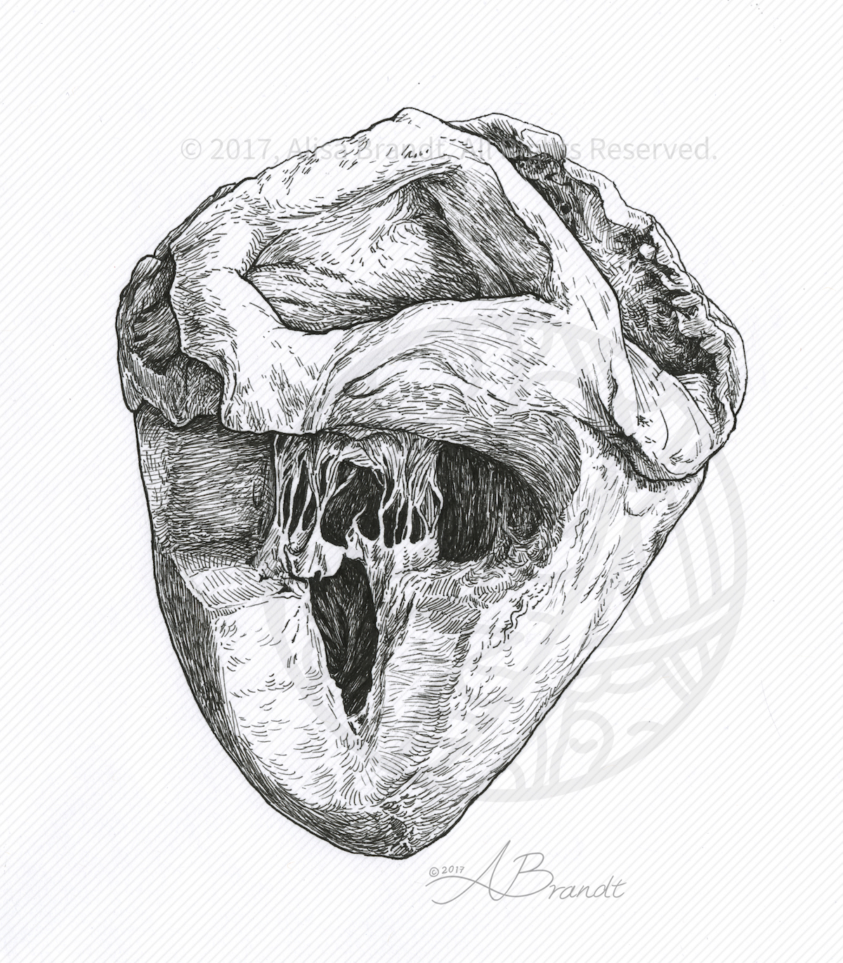
Pen and ink sketch from direct observation of a plastinated heart specimen. Created during the Medical and Biological Illustration graduate program at Johns Hopkins University School of Medicine.
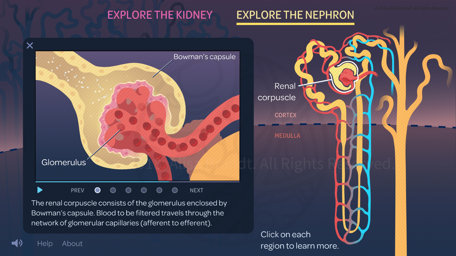
Screenshot of a 2D interactive web module prototype, for teaching introductory physiology of the nephron of the kidney. Created during the Medical and Biological Illustration graduate program at Johns Hopkins University School of Medicine. View interactive media
Anatomical sketch from direct observation of the mesentery and mesenteric vessels of the small intestine. Created during the Medical and Biological Illustration graduate program at Johns Hopkins University School of Medicine.

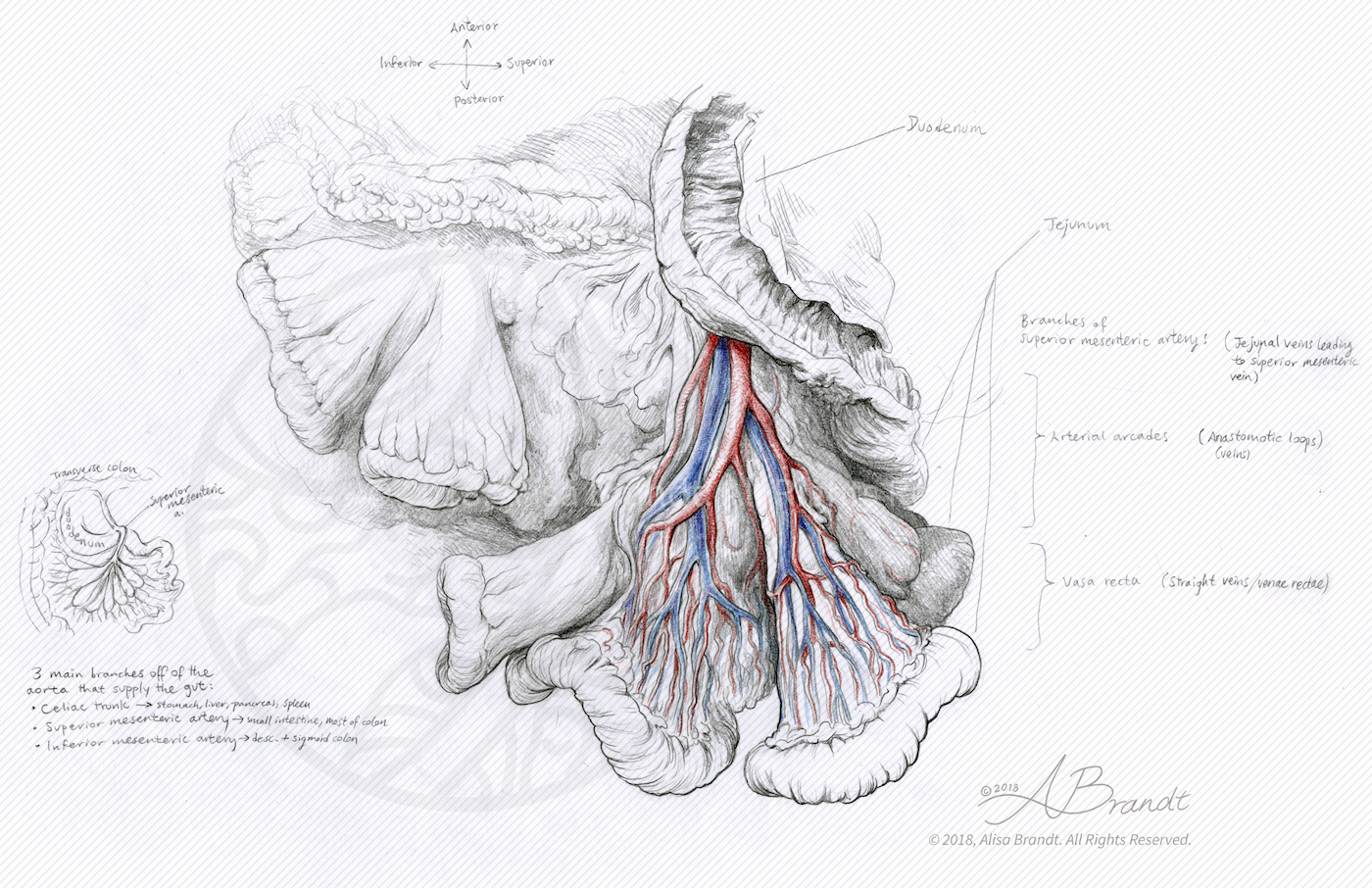
Anatomical sketch from direct observation of the kidneys and abdominal vessels. Created during the Medical and Biological Illustration graduate program at Johns Hopkins University School of Medicine.
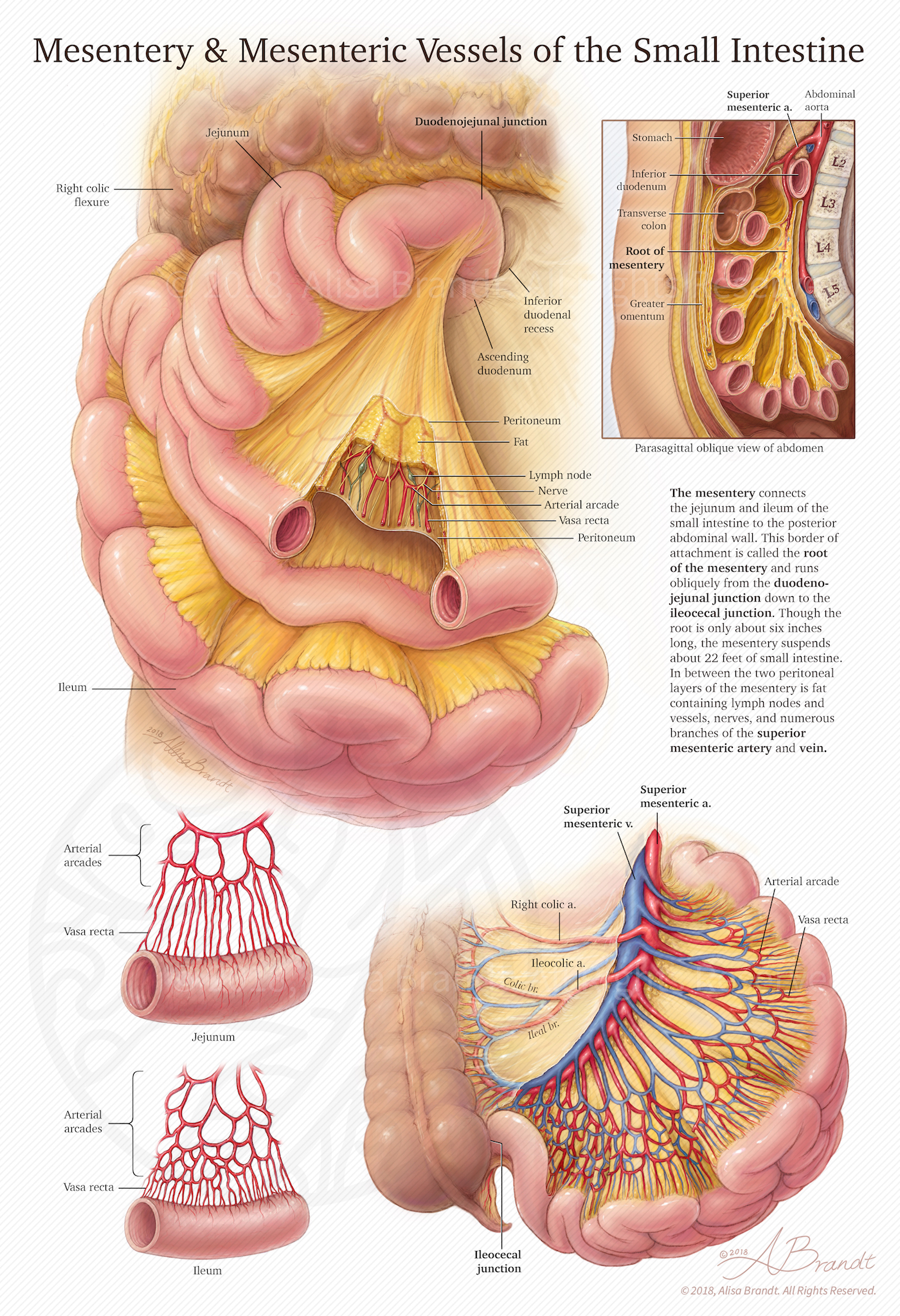
Anatomical plate about the mesentery and mesenteric vessels of the small intestine, created during the Medical and Biological Illustration graduate program at Johns Hopkins University School of Medicine
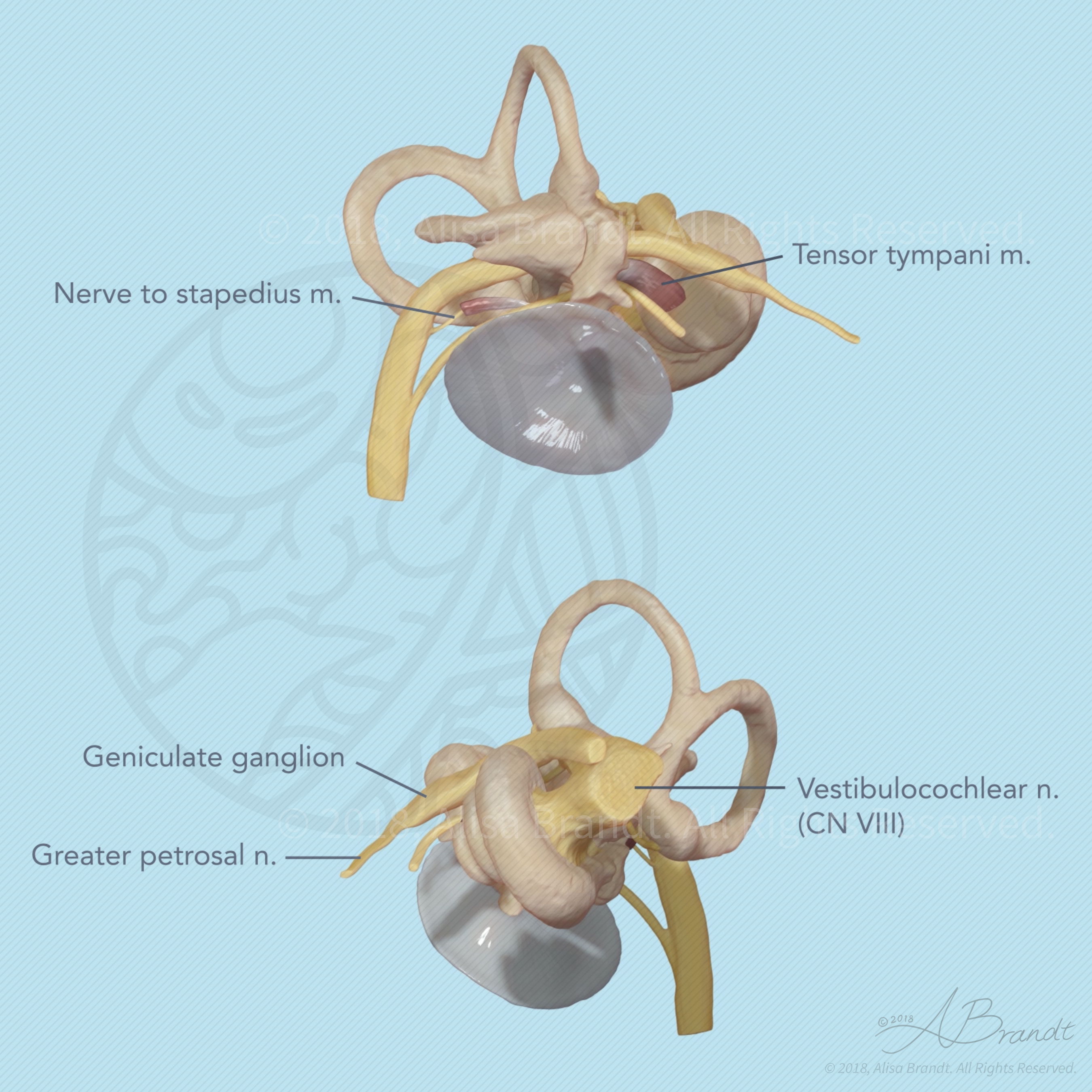
Still images from a turntable movie (click here) of the inner and middle ear sculpted using ZBrush, based on MRM (magnetic resonance microscopy) data. The anatomy of the inner and middle ear is presented with a particular focus on the nerves of the area. The MRM data (a series of TIFF images) was prepared in OsiriX and a 3D surface was exported to OBJ format for sculpting in ZBrush. Data