Red-capped cardinals poster

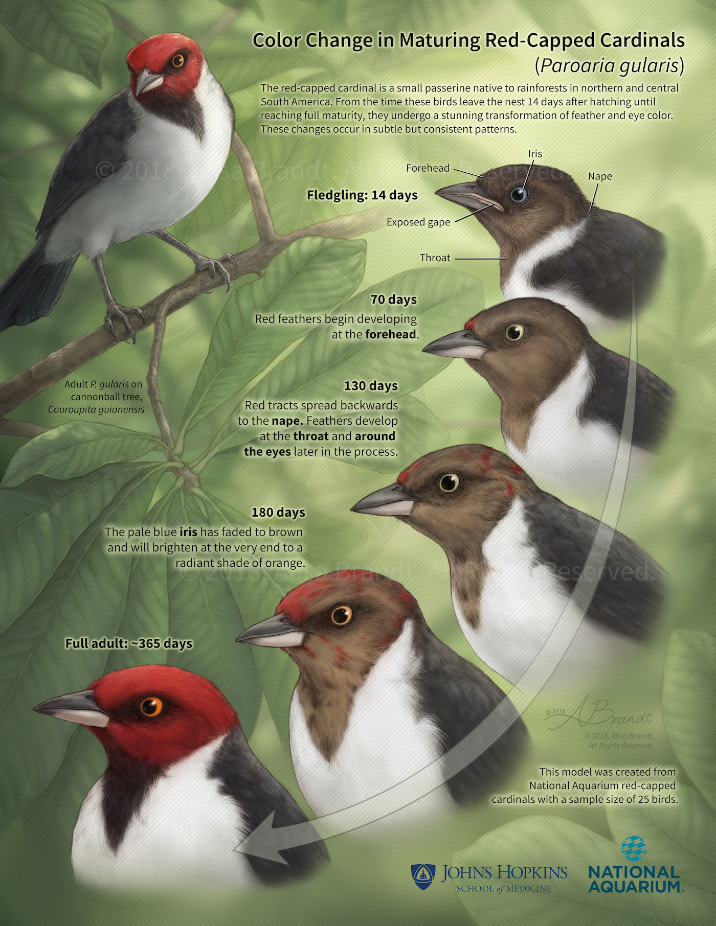
Color Change in Maturing Red-Capped Cardinals

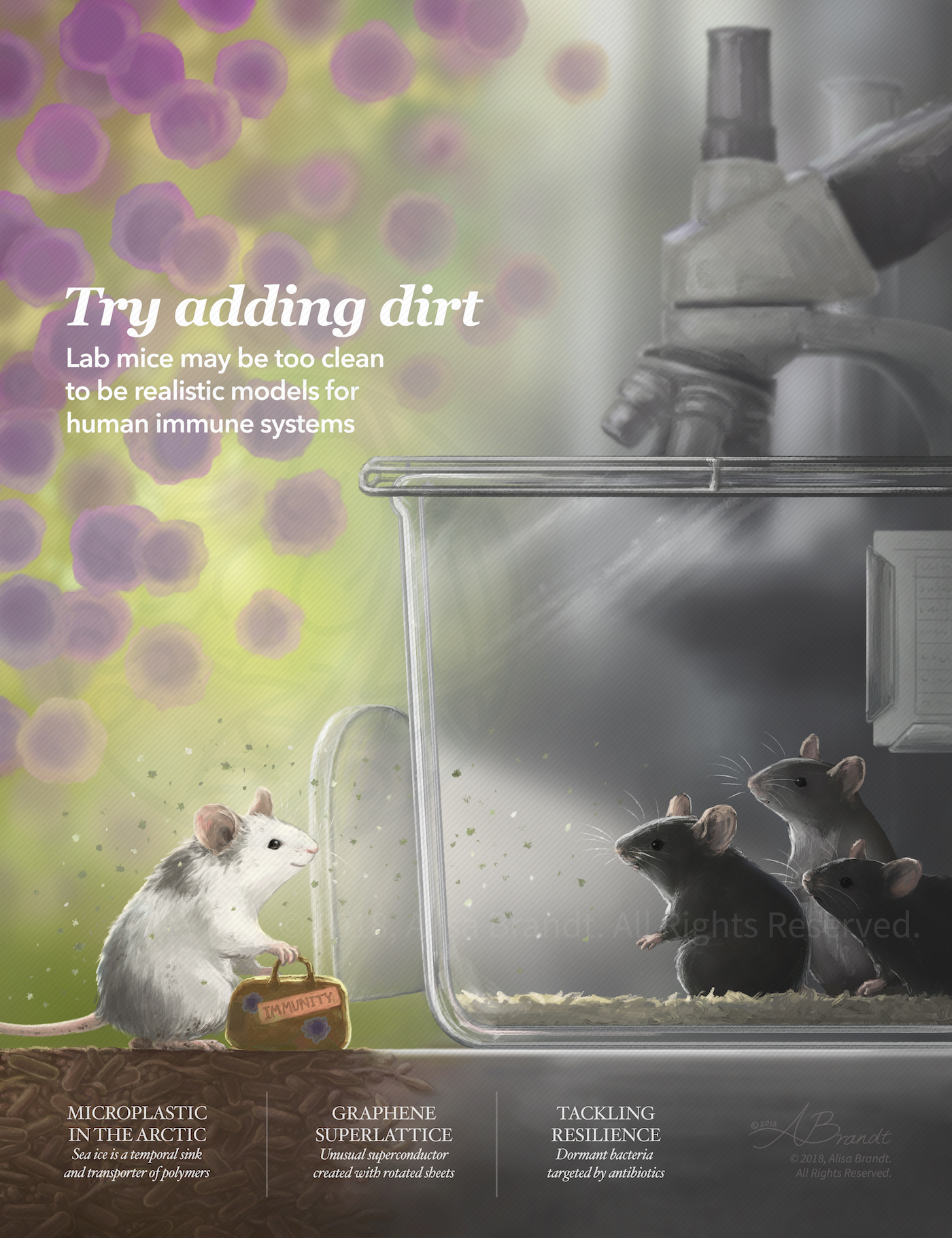
Illustration created for the National Aquarium in Baltimore, Maryland, during the Medical and Biological Illustration graduate program at Johns Hopkins University School of Medicine. This poster communicates the typical progression of color change in red-capped cardinals as they mature from fledgling to adult.

Still images from inner and middle ear turntable movie

The Inner and Middle Ear (stills)

Still images from a turntable movie (click here) of the inner and middle ear sculpted using ZBrush, based on MRM (magnetic resonance microscopy) data. The anatomy of the inner and middle ear is presented with a particular focus on the nerves of the area. The MRM data (a series of TIFF images) was prepared in OsiriX and a 3D surface was exported to OBJ format for sculpting in ZBrush. Data

Bumblebees poster

Nectar and Pollen Gathering of Bumblebees

Educational poster about bumblebees and their adaptations for nectar and pollen gathering, combining graphite and Photoshop. Created during the Medical and Biological Illustration graduate program at Johns Hopkins University School of Medicine.

Laparoscopic sigmoid colectomy surgical illustration

Laparoscopic Sigmoid Colectomy

Laparoscopic sigmoid colectomy surgical illustration designed for a surgical atlas. Created during the Surgical Illustration course in the Medical and Biological Illustration graduate program at Johns Hopkins University School of Medicine.

Molecular immunotherapy poster

Immunotherapy to Fight Cancer

Immune checkpoint blocking therapies, or ICBs, help fight cancer by inhibiting the down-regulation of effector T cell activity. Here, two CD8+ T cells (in blue) approach a cancer cell (in brown), while ICB antibodies flow in from the upper right to block interaction between PD-1 and PDL-1. Peptide fragments (in red) are being presented by the cancer cell on its surface in HLA-I (Human Leukocyte Antigen) molecules, which make up

Neuroanatomical illustration

Anatomy of the Floor of the Lateral Ventricle

Illustration created during the Neuroanatomy course in the Medical and Biological Illustration graduate program at the Department of Art as Applied to Medicine, Johns Hopkins University School of Medicine. This illustration teaches the anatomy of the human brain around the lateral ventricle, a fluid-filled space that extends into the temporal lobe. The brain has been cut in several planes to expose the floor of the left lateral ventricle and the

Mock journal cover editorial illustration

Mock Journal Cover

Editorial illustration created during the Medical and Biological Illustration graduate program at Johns Hopkins University School of Medicine. The article chosen to be illustrated was: Willyard, C. 2018. Send in the germs: Lab mice are usually kept squeaky clean, but some immunologists think a dose of dirt could make them more useful for science. Nature, 556: 16-18.

Scroll UpScroll Up
error: Content is protected !!